Waves, Wires, and Vision: How Cells Pull Together to Build the Eye
08-12-2025
New research from Purdue University reveals how developing eye tissue transforms itself from a flat sheet into a precisely curved structure, offering insight into how mechanical forces shape organs — and what might go wrong when those forces fail.
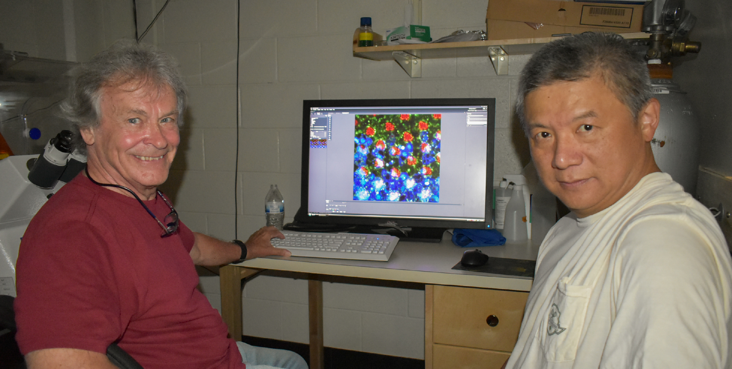
Published in iScience, the study from Purdue biological sciences professors Donald Ready and Henry Chang titled Drosophila photoreceptor tethering by a laminin-Eys scaffold, explores how specific cells coordinate to sculpt a complex, dome-like shape using a combination of structural proteins, internal tension, and wave-like calcium signals.
The findings help explain a fundamental process in developmental biology: how tissue shape is generated and maintained at the cellular level. And because similar structural systems are found throughout the body — including in human tissues — the implications extend far beyond vision.
"This work shows how contractile forces inside developing tissue are tightly coordinated to create a specific shape," said Chang. "In this case, we studied how cells use internal actin fibers and calcium signals to reshape the eye from a flat layer into a curved structure that can support wide-angle vision."
The transformation is guided by a specialized scaffold called the extracellular matrix (ECM), a network of proteins that supports and connects cells. The researchers discovered that this matrix is not just passive support — it’s dynamically assembled in coordination with the tissue itself. Specific ECM proteins act as anchor points for contractile fibers within the cells, allowing the tissue to pull itself into a new shape.
"Think of it like architectural engineering," said Ready. "The matrix forms a kind of internal suspension system, and the cells apply tension through it. It’s a mechanical solution to a biological challenge."
To track how this happens during development, the team used high-resolution imaging and genetic tools to observe how ECM structures form and how calcium signals move through the tissue like waves. These calcium waves help synchronize contractions across the tissue, reducing its surface area and producing the final curvature.
The analogy is not just visual. The researchers compare the tension-based structure to the roof of Madison Square Garden, where steel cables connect to a central ring to evenly distribute force. Similarly, in the eye, contractile fibers link to ECM "grommets" at the tissue floor, transmitting tension through a central ring-like structure.
The research also offers new clues to the causes of vision loss. One of the ECM proteins in this system — called Eys — is essential for keeping the eye’s light-sensing cells in perfect alignment. In humans, mutations in the gene that produces Eys are the most common cause of retinitis pigmentosa, a degenerative disease that leads to blindness. Until now, it was unclear why these mutations disrupted vision.
"Beyond basic biology, the work also has clinical relevance," Ready said. "Our study shows Eys plays a role in organizing other ECMs. Mutations in human Eys cause neurodegeneration, and while they are the most common cause of retinitis pigmentosa, it has not been known why they cause the disease. This work reveals a novel interaction between Eys and another ECM protein, laminin. Together, they build a central core that supports the tension needed to keep photoreceptors aligned."
Because the same molecular components are found across many species, this model system offers a powerful way to investigate Eys function and its role in disease — without the limits of studying only human tissue.The study was supported by a Purdue Research Refresh Award and funding from the National Institutes of Health. Imaging was made possible in part through an NIH Shared Instrumentation Grant.
"This model has helped us see how structure and function come together at the cellular level," said Ready. "It’s a detailed look at how tissues form — and a step toward understanding how we might one day repair them."
About the Department of Biological Sciences at Purdue University
The Department of Biological Sciences is the largest life sciences department at Purdue University. As part of Purdue One Health, we are dedicated to pioneering scientific discoveries and transformative education at the cutting edge of innovation. From molecules to cells, from tissues to organisms, from populations to ecosystems - we bring together multiple perspectives, integrating across biological scales to advance our understanding of life and tackle the world’s most pressing challenges. Learn more at bio.purdue.edu/.
Writer: Alisha Willett, amwillet@purdue.edu
Source: Henry Chang, hcchang@purdue.edu and Donald Ready, dready@purdue.edu