Molecular Basis of Heterotrimeric G Protein Function
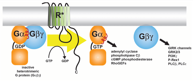
Signal transduction from β-adrenergic receptors to adenylyl cyclase via heterotrimeric G proteins is one of the classic paradigms of hormone action, serving as a model system for the hundreds of G protein-coupled receptors (GPCRs) found in the human genome. Over the last two decades, monumental strides were made in understanding the molecular basis for how this GPCR-mediated signaling occurs. The Tesmer lab has played a key role in this process by using the power of X-ray crystallography, and more recently cryo-electron microscopy, to illuminate the mechanisms of these essential cellular processes at atomic resolution. While working as a post-doctoral fellow with Dr. Stephen Sprang, Dr. Tesmer reported the crystal structures of Gαs alone and in complex with the catalytic domains of adenylyl cyclase (1), which was the first atomic structure of a Gα-effector complex. More recently the Tesmer Lab has focused on effector enzymes responsive to Gαq and Gβγ subunits, such as GRK2 (2), PLCβ (3), and P-Rex1(4), respectively.
Publications
-
Tesmer JJ, Sunahara RK, Gilman AG, Sprang SR. Crystal structure of the catalytic domains of adenylyl cyclase in a complex with Gsα·GTPγS. Science. 1997 278:1907-16. PMID: 9417641.
-
Tesmer VM, Kawano T, Shankaranarayanan A, Kozasa T, Tesmer JJG: Snapshot of activated G proteins at the membrane: the Gαq-GRK2-Gβγ complex. Science 2005, 310: 1686-1690. PMID: 16339447.
- Lyon AM, Dutta S, Boguth CA, Skiniotis G, Tesmer JJ. Full-length Gαq-phospholipase C-β3 structure reveals interfaces of the C-terminal coiled-coil domain. Nat Struct Mol Biol. 2013 20:355-62. PMC3594540.
-
Cash JN, Urata S, Li S, Ravala S, Avramova L, Gutkind JS, Tesmer JJG, Cianfrocco MA: Cryo-electron microscopy structure and analyses of the P-Rex1–Gβγ complex signaling scaffold. Sci Advances 2019, 5: eaax8855. PMC6795519